A new method to reveal the molecular landscapes of photosynthetic membranes inside green algae cells
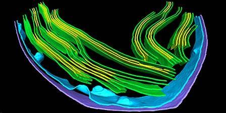
A collaboration with Max Planck Institutes in Germany has led to a new visualization approach that produces a topological view of these native membranes.

Scientific Achievement
The latest advances in cryo-electron tomography were used to image photosynthetic protein complexes embedded within native thylakoid membranes inside the cell.
Significance and Impact
This high-resolution imaging revealed how the intricate network of thylakoid membranes sorts protein complexes into distinct regions to tune the photosynthetic reactions. A surprising discovery shows how large Photosystem II supercomplexes may be able to move fluidly through the crowded membranes, maintaining efficient capture of light energy and transfer of electrons to other complexes.
Research Details
- A new visualization approach called a ‘membranogram’ projects tomographic images of the proteins onto the surface of the segmented membrane, producing a molecular view of native membrane topology.
- PSII were mostly found in the appressed region, whereas PSI, ATP synthase and ribosomes were restricted to the non-appressed region, and Cytb6f were found with equal abundance in both regions.
- PSII supercomplexes randomly overwrap between appressed membranes, apparently not maintaining stacking.
Related people: Wojciech Wietrzynski, Miroslava Schaffer, Dimitry Tegunov, Sahradha Albert, Atsuko Kanazawa, Jürgen M Plitzko, Wolfgang Baumeister, Benjamin D Engel (CA)
DOI: 10.7554/eLife.53740
This work was partially funded by the US Department of Energy, Office of Basic Energy Sciences. Contributions from Dr. Kanazawa, from the MSU-DOE Plant Research Laboratory, include: Data curation, Formal analysis, Investigation, and 77K measurements


